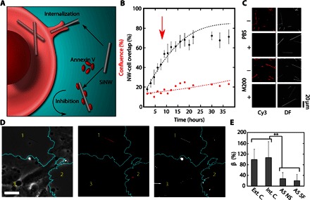
Fig. 5. A5 as a phagocytosis inhibitor of SiNWs.

(A) Schematic illustration showing the inhibition mechanism of A5. Unlike inhibitors that target the cell directly, the nonspecific binding of A5 to the negatively charged SiNW surface can screen the SiNW from uptake. (B) A5 inhibitor study, showing a reduced nanowire-cell overlap (black dots) as compared to the expected internalization (black line) (red arrow indicates dosage time). (C) Fluorescent and DF micrographs showing the level of A5-Cy3 absorption onto SiNWs in the presence (+) and absence (−) of serum in PBS and M200 solutions, indicating that the ability of A5 to bind with SiNWs is restricted by the presence of serum proteins. (D) Example micrograph showing the same region in the PC (left), DF (middle), and Cy3 (right, artificial color) channels, indicating that SiNW surfaces modified with Cy3-A5 (1 and 2) are excluded from the cell, whereas the internal controls (3) are endocytosed (artificial cell outline, teal) (scale bar, 30 μm). (E) SiNW-cell overlap in HUVECs after 24 hours of SiNWs with nonspecifically bound A5-Cy3 (A5 NS) and surface-functionalized A5-Cy3 (A5 SF), relative to an external (Ext. C.) and internal (Int. C.) control of unmodified SiNWs (β given as a percentage of external control) (**P < 0.01). DF and Cy3 were background-subtracted using a Gaussian spatial filter uniformly applied to each image.
