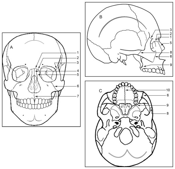
Fig 1.
Schematic representation of the sutures used in the study: A, frontal; B, lateral; C, axial views. 1, Frontonasal; 2, frontomaxillary; 3, frontozygomatic; 4, internasal; 5, nasomaxillary; 6, zygomaticomaxillary; 7, intermaxillary; 8, temporozygomatic; 9, pterygomaxillary; 10, midpalatal suture.

