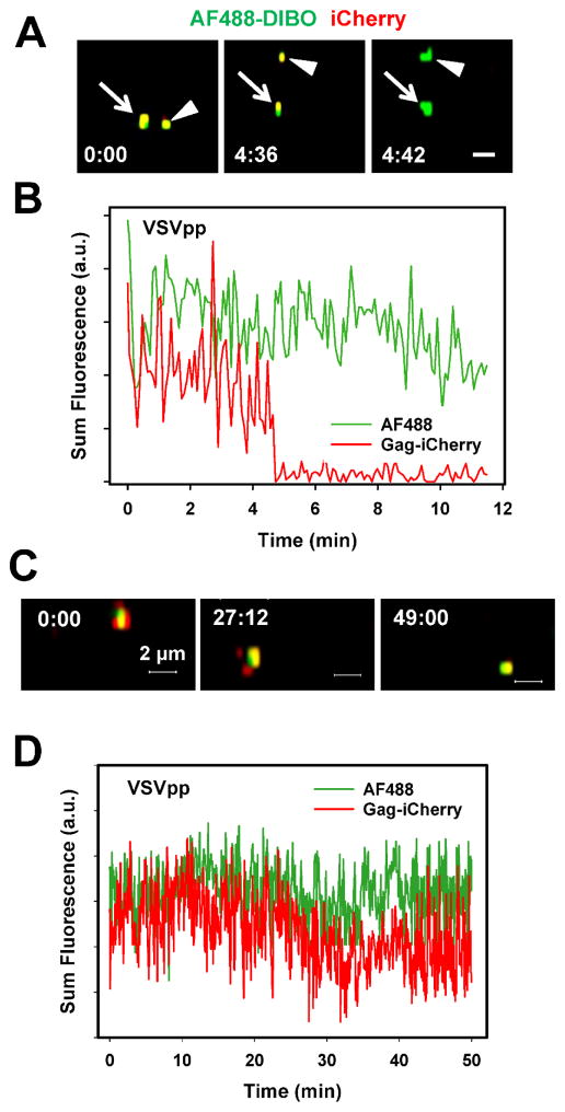
Figure 4. Fusion of single VSVpp co-labeled with Alexa488-DIBO (green) and Gag-imCherry (red).
(A) Images of two double-labeled VSVpp fusing with endosomes of CV-1-derived cells nearly at the same time, as manifested in change from colocalized yellow color to green color. Scale bar 2 μm. (B) Sum fluorescence intensity profiles for the lower particle in panel A obtained by single particle tracking. (C, D) images and fluorescence profiles for a control double-labeled particle that did not undergo fusion.

