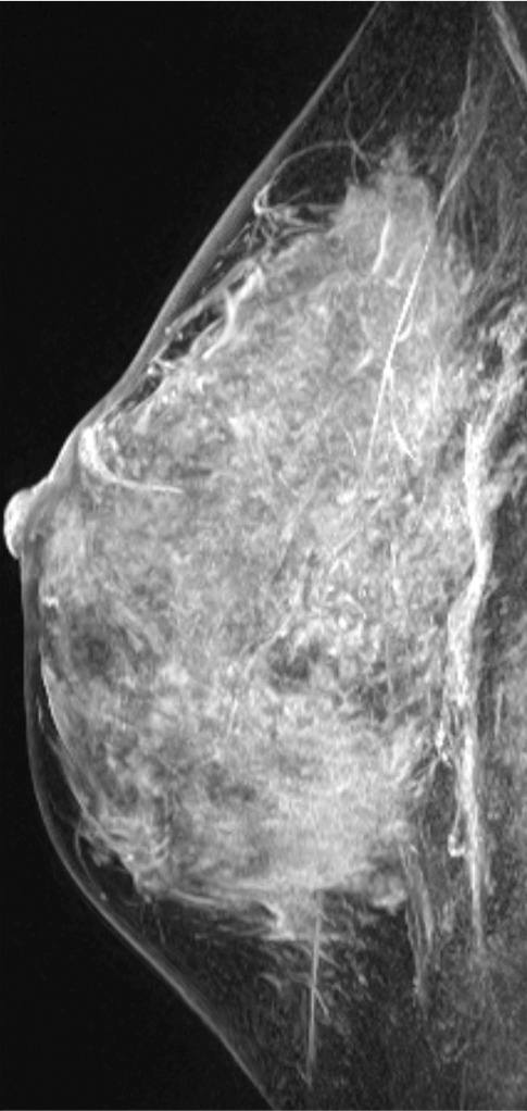
Figure 11c.

Interval cancer that was DBT occult at routine screening of a 54-year-old woman. (a) MLO DBT image obtained at screening was interpreted as normal. Five months after screening, the patient presented with diffuse pain and thickening in the right breast. (b) Diagnostic MLO DBT image (b) and targeted US images (not shown) were also interpreted as negative. Given the patient’s symptoms, MR imaging was performed. (c) Contrast-enhanced MIP MR image shows diffuse, asymmetric, nonmasslike enhancement. (d) MIP MR image of the normal contralateral breast is shown for comparison. (e) Axial postcontrast fat-suppressed T1-weighted MR image shows global asymmetric nonmasslike enhancement of the right breast relative to the left breast. Biopsy revealed diffuse micropapillary ductal carcinoma in situ with areas of microinvasion. The patient’s clinical symptoms should always guide management, and not all cancers may be seen at DBT.
