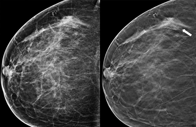
Figure 2a.
Cancer seen only at DBT at routine screening of a 46-year-old woman. (a, b) CC DM (left) and CC DBT (right) images (a) and MLO DM (left) and MLO DBT (right) images (b) demonstrate that an area of subtle architectural distortion (arrow) is visible in the upper outer quadrant on the DBT images only. (c) Ultrasonographic (US) image shows an ill-defined hypoechoic mass. (d) Sagittal magnetic resonance (MR) subtraction image shows a region of architectural distortion with nonmasslike enhancement. Biopsy revealed a 6-cm invasive lobular carcinoma.

