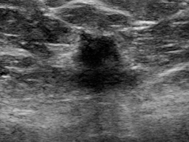
Figure 3c.

Cancer seen better on CC views than MLO views at both DM and DBT at routine screening of a 78-year-old woman. (a, b) CC DBT image (a) shows an irregular mass with spiculation (arrow in a and b) that was also seen on a CC DM view (not shown). Using the quasi–three-dimensional localization available with the CC DBT view, the lesion was triangulated to the medial and mid breast on the MLO DBT view (b), where it is subtle yet visible. The lesion was not visible on an MLO DM view (not shown) because of superimposed tissue and lack of any detectable distortion. (c) US image shows an irregular hypoechoic mass. Biopsy revealed intermediate-grade invasive ductal carcinoma.
