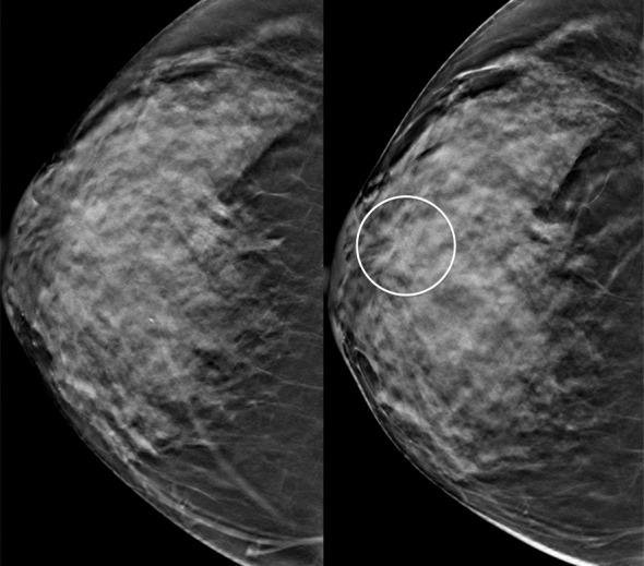
Figure 5a.

Interval cancer at DBT. (a) CC DBT image obtained at routine screening (left) was interpreted as negative. The patient returned 6 months later with a palpable mass in the subareolar location, which was then detected at DBT on the CC view only (right) as an area of subtle distortion (circle). (b) US image shows an irregular mass with calcifications. Biopsy revealed intermediate-grade invasive ductal carcinoma with associated ductal carcinoma in situ. Dense breast tissue may cause perceptual errors at both DM and DBT because cancers may be obscured by overlying complex tissue patterns. Although DBT has improved cancer detection even in dense breasts, some cancers will be subtle and difficult or impossible to detect.
