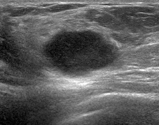
Figure 7c.

Interval cancer in a 49-year-old woman. (a) MLO DBT image obtained at routine screening shows a possible area of architectural distortion (arrow) in the superior breast that was seen on this view only. (b) MLO DM spot compression image obtained at recall was interpreted as negative. An enlarged lymph node at the edge of the field of view (arrow) was not noted. (c, d) US (c) and sagittal contrast-enhanced T1-weighted MR (d) images obtained 8 months later, when the patient presented with an axillary tail mass, show a corresponding irregular mass (arrow in d). Biopsy revealed poorly differentiated invasive ductal carcinoma. Both a cognitive error (not recognizing the subtle signs of malignancy on the recalled diagnostic study) and a perceptual error (not recognizing the enlarged lymph node at the corner of the image) contributed to a missed opportunity to detect malignancy earlier.
