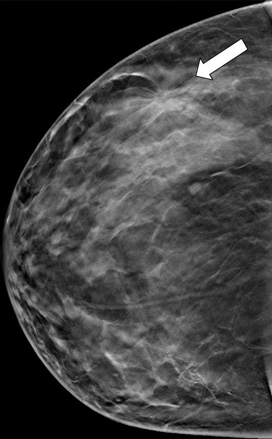
Figure 9b.

Suspicious finding attributed to postoperative change in a 51-year-old woman with a history of a benign remote surgical biopsy in the upper outer quadrant who presented for routine screening. (a) CC DBT image shows architectural distortion in the right lateral breast, a finding that was incorrectly attributed to the site of the patient’s remote biopsy (which was in the upper outer breast). (b) CC DBT image obtained at screening 1 year later again demonstrates increasing architectural distortion (arrow) in the lateral breast. The finding was correctly triangulated on the single-view CC DBT as in the lower breast (not at the site of the prior surgery in the upper outer breast). (c) With careful attention to the quasi–three-dimensional information obtained from the CC DBT view, the subtle lesion (arrow) can now be seen on an MLO DBT view. (d) US image reveals a hypoechoic spiculated mass. (e) Sagittal maximum intensity projection (MIP) MR image shows an enhancing spiculated mass (arrow) in the central breast. Biopsy revealed intermediate-grade invasive ductal carcinoma. The site of a patient’s surgical scar should always be confirmed before attributing a finding to postbiopsy changes.
