Figure 5.
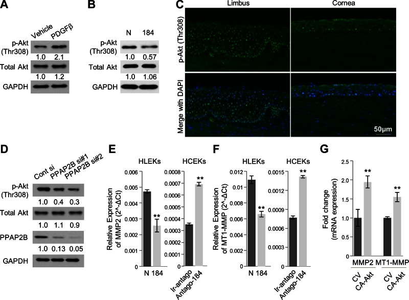
miR-184 modulates Akt activity and its downstream gene expression. A) Immunoblotting of phospho-Akt (T308) and total Akt expression in HCEKs after treatment of PDGF-β. B) Immunoblotting of phospho-Akt (T308) and total Akt expression in HLEKs after transfection of precursor miRNA negative control or precursor miR-184. Phospho-Akt was markedly decreased in HLEKs expressing miR-184 compared with controls. C) Tissue immunofluorescence staining with p-Akt antibody in the frozen section of human limbal and corneal epithelium. Phospho-akt was strongly localized in the nucleus of limbal epithelium compared with corneal epithelium. D) Both phospho-Akt (T308) and total Akt protein levels were determined in HLEKs after depletion of PPAP2B using siRNAs. E) Real-time qPCR analysis of MMP2 levels in HLEKs and HCEKs after overexpression or knockdown of miR-184, respectively. F) MT1-MMP levels were determined by qPCR in HLEKs and HCEKs after overexpression or knockdown of miR-184, respectively. G) Real-time qPCR analysis of MMP2 and MT1-MMP in HCEKs followed by transient transfection with either control vector (CV) or a constitutively active Akt (CA-Akt). Expression levels were calculated relative to GAPDH mRNA.

