Figure 5.
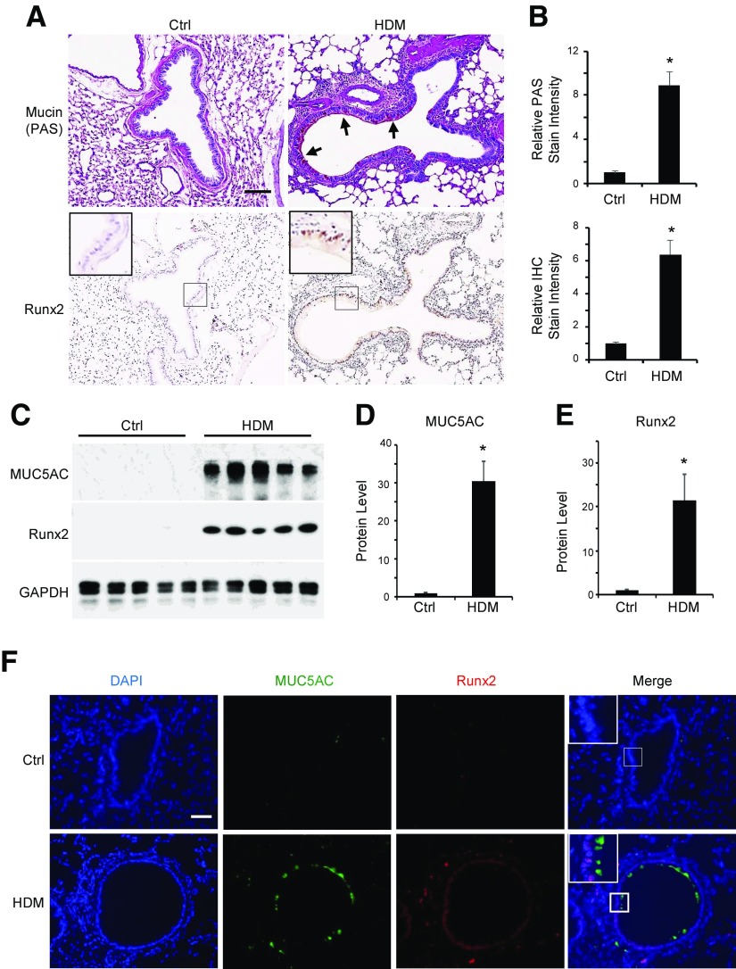
Runx2 was induced in HDM-induced goblet cell differentiation in mouse lungs. Mice were sensitized and challenged with HDM as described in Materials and Methods. A) Mouse lungs were harvested and sectioned, followed by PAS staining (top) for mucus production and IHC staining for Runx2 expression (bottom). Arrows point to PAS-stained mucin. B) Quantification of mucin production (top) and Runx2 expression (bottom) shown in (A) by normalizing to staining density in control groups (ctrl). *P < 0.01 compared to control (n = 5). C–E) Runx2 correlated with mucin 5AC expression in HDM-challenged mouse lungs. Mouse lungs were collected 3 d after last HDM challenge (PBS as control). Western blot analysis (C) was performed to examine Runx2 and mucin 5AC expression. Mucin 5AC (D) and Runx2 (E) protein levels shown in C were quantified by normalizing to GAPDH level, respectively. *P < 0.01 compared to control (n = 5). F) HDM-induced Runx2 and mucin were co-localized in same pulmonary epithelium. Immunofluorescence staining was performed to display mucin 5AC and Runx2 expression. DAPI stains nuclei. Scale bars, 100 µm.

