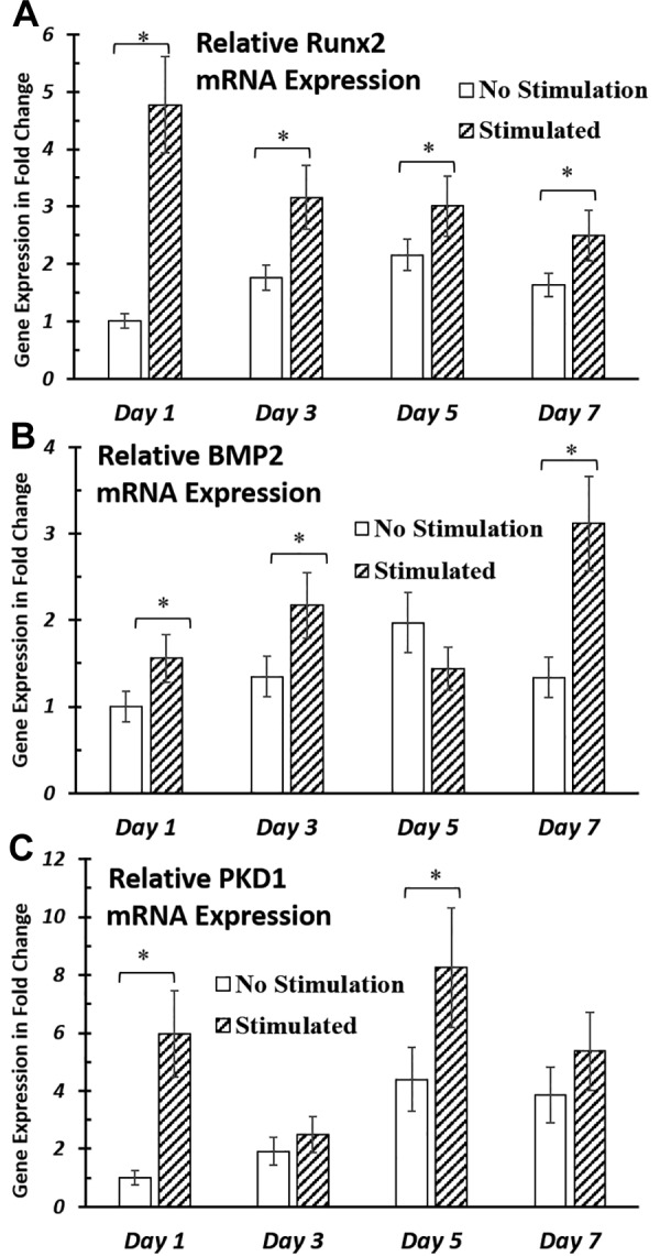
Figure 4.

Gene expression of osteogenic markers and primary cilia structural proteins after up to 7 d of EFS. A, B) EFS significantly improved gene expression of osteogenic markers Runx2 (A) and BMP-2 (B) in hASCs during up to 7 d of stimulation. C) mRNA expression of primary cilia structural protein, PC1, was up-regulated by EFS at timepoints d 1 and 7. hASC-seeded IDEs were cultured in ODM and stimulated with 1 V/cm electric field at 1 Hz for 4 h/d. RNA was extracted and analyzed at the end of electrical stimulations for respective timepoints. mRNA expressions were normalized to nonstimulated samples at d 1. Error bars = sem, Student’s t test compared stimulated samples with nonstimulated control samples at each timepoint. *P < 0.05.
