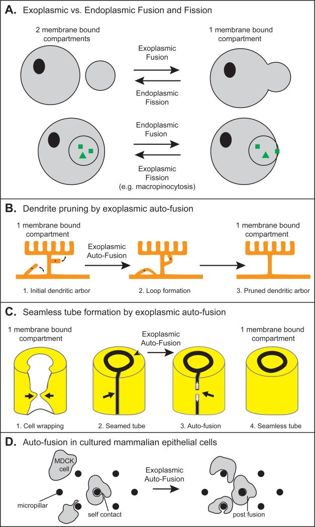
Figure 1. Fission, Fusion and Auto-fusion.
(A) Comparison of different types of fission and fusion, adapted from [7]. Cytoplasmic domains are marked with grey coloring. (B-D) Examples of exoplasmic auto-fusion maintaining one membrane bound compartment. (B) A C. elegans arborized dendrite (“menorah”) has two ectopic branches. Auto-fusion induces loop formation to prune the ectopic branches and maintain only the appropriate right-angled branches [4]. (C) The three steps of seamless tube formation [5, 6, 79]. (1) Cell wrapping. White, apical surface; yellow, basal surface. The arrows show the path of cell wrapping. (2) Formation of a seamed tube. An auto-cellular junction (arrow) maintains the cell in the shape of a tube. A ring junction (arrow-head) seals the cell with its neighbors (not shown) to continue the tube. (3) Proposed model of Auto-fusion. A fusion pore forms (black arrow) and extends (white arrows) to form a seamless tube (4). (D) Model of auto-fusion in cultured mammalian MDCK cells [91].

