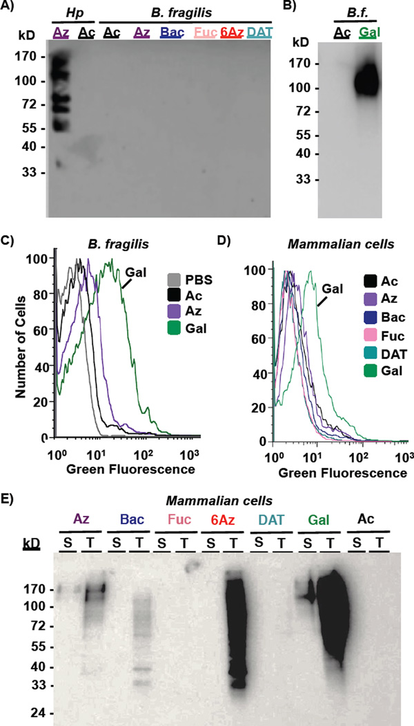
Figure 5.
Metabolic incorporation of azide-containing rare bacterial monosaccharide analogs onto symbiotic Bacteroides fragilis and mammalian Madin Darby Canine Kidney (MDCK) cells. A–C) B. fragilis were grown in media supplemented with 1 mM Ac4GlcNAz (2, Az), Ac2Bac-diNAz (5, Bac), Ac3FucNAz (4, Fuc), Ac36AzGlcNAc (3, 6Az), Ac2DATDG-diNAz (6, DAT), or the azide-free control sugar Ac4GlcNAc (1, Ac) for 2 days, then probed for the presence of azide-labeled glycans on cells or within lysates by reaction with 250 µM Phos-FLAG and subsequent detection with anti-FLAG antibody via Western blot (A, B) or by reaction with DIBO-488 (20 µM, 1 hour) followed by flow cytometry (C). D, E) MDCK cells were cultured with 0.25 mM azidosugar-supplemented media for 3 days, then probed for the presence of azide-labeled glycans using Phos-FLAG and anti-FLAG antibody. D) Analysis of intact MDCK was performed by flow cytometry with anti-FLAG-FITC. E) Incorporation of azides into MDCK surface-accessible (“S”) versus total (“T”) glycoproteins was probed by reacting intact cells or cell lysates, respectively, with Phos-FLAG, then analyzing samples via Western blot analysis. Data are representative of replicate experiments (n≥3).

