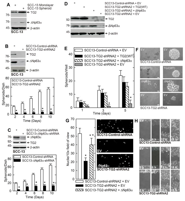
Fig. 1. TG2 and ΔNp63α are enriched in ECS cells, and required for ECS cell survival.
A ECS cells have elevated TG2 and ΔNp63α. SCC-13 cells (40,000 per well) were grown in spheroid medium in attached (monolayer) or non-attached (spheroid, ECS cell) conditions. At 10 d lysates were electrophoresed for detection of the indicated epitopes. B SCC13-Control-shRNA and SCC13-TG2-shRNA2 cells were grown in spheroid culture for 10 d prior to counting of spheroid number and extract preparation for detection of TG2 and ΔNp63α. C SCC13-Control-shRNA and SCC13-ΔNp63α-shRNA cells were grown in spheroid culture for 10 d prior to counting of spheroid number and extract preparation for detection of TG2 and ΔNp63α. D SCC13-Control-shRNA or SCC13-TG2-shRNA2 cells were double-electroporated with empty vector (EV) or expression vectors encoding TG2 or ΔNp63α and after 5 d the indicated proteins were detected. E/F The indicated cell lines were electroporated with EV, TG2(WT) or ΔNp63α and grown as spheroids for 5 d prior to counting of spheroids and recording of images. G The indicated cell lines was electroporated with the indicated plasmid and after 48 h seeded onto a matrigel-coated membrane in a Millicell chamber for invasion assay. After 20 h the membrane was removed, fixed, and DAPI-stained for nuclei counting. H Cells were electroporated with the indicated plasmid, plated at high density to form confluent monolayers, the monolayers were scratched with a 10 μl pipette tip to create a wound and wound closure was monitored with time. The plotted values are mean ± SEM and asterisks indicate significant change compared to control, n = 3, p < 0.005. The double asterisk in panel G indicates a significant change compared to the single asterisk group, n = 3, p < 0.005.

