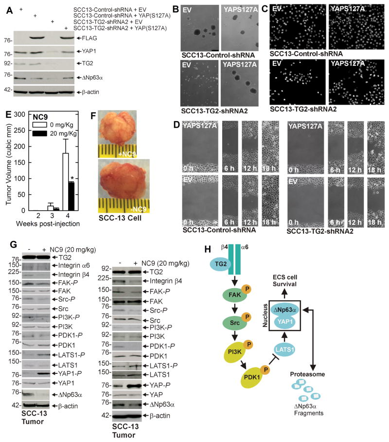
Fig. 4. YAP1 enhances ECS cell survival and TG2 inhibitor suppresses tumor formation.
A SCC13-Control-shRNA and SCC13-TG2-shRNA2 cells were double electroporated with empty vector (EV) or YAP(S127A)-encoding vector and at 48 h post-electroporation lysates were collected for immunoblot. B/C/D The indicated cell lines were electroporated with EV or YAP(S127A)-encoding vector and then plated for spheroid formation, invasion and migration assay. E/F ECS cells (100,000 cells derived from SCC-13) were injected into each front flank in NSG mice. At 1 d post injection, NC9 was delivered by intraperitoneal injection, three times per week on alternate days, of 200 μl of a 2 mg/ml stock (20 mg/kg body weight). Images represent appearance and size of typical control and NC9 treated tumors harvested on week four. The plotted values are mean ± SEM and asterisks indicate significant change compared to control, n = 5 mice (10 tumors), p < 0.001. G Tumors were harvested at 4 wk and extracts were prepared for assay of indicated proteins. Blots from two representative tumors are shown. H Proposed TG2 signaling scheme. TG2 interacts with α6/β4 integrin to enhance integrin (FAK/Src) signaling which increases PI3K/PDK1 activity and PDK1 suppresses activity in the Hippo signaling cascade (LATS1). This leads to reduced LATS1 phosphorylation of YAP1 which then interacts with and stabilizes ΔNp63α. This signaling pathway enhances ECS cell survival, invasion and migration. Loss of TG2 function reduces YAP1 which leads to degradation of ΔNp63α and reduced ECS cell survival, invasion and migration. Similar results were observed in extract prepared from additional tumors.

