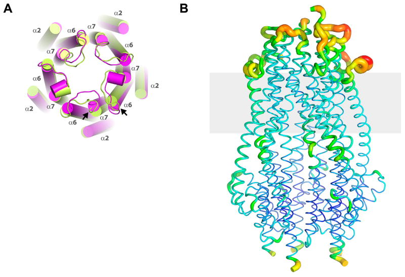
Extended Data Figure 3. Helical shifts and overall flexibility in the ExbB pentamer.
A. Two pentamers were observed per asymmetric unit within the crystal structure. Shown here is pentamer 1 (green) aligned with pentamer 2 (magenta), illustrating slight shifts in a number of the helices (cylinders) between the two pentamers, with the largest shifts indicated by the black arrows. Further, the loops connecting α6 and α7 also show variability between monomers and pentamers. B. The TonB subcomplex (ExbB/ExbDΔperi) showing a B-factor putty representation with values ranging from the most ordered in blue to the most disordered in red.

