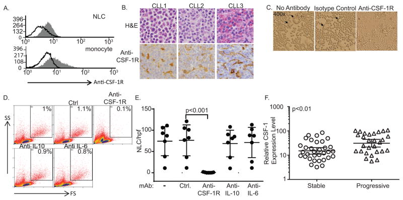
Figure 1.
CSF-1R is required for nurse-like cell survival. (A) Cultured PBMC from CLL patients were stained with an isotype control (open histogram) or anti-CSF-1R (gray histogram). NLC, identified by forward and side scatter, were gated and CSF-1R expression determined by flow cytometry. A representative example is shown (n=3). (B) Immunohistochemical staining for CSF-1R was performed in paraffin-embedded CLL (n=43). Staining of CLL-associated macrophages is appreciated in the representative lymph nodes (CLL1, CLL2) and spleen (CLL3) shown. (Original magnification 400x) (C–E) PBMC from CLL patients (n=7) were cultured in the presence or absence of an isotype control or antagonistic anti-CSF-1R, anti-IL-10, or anti-IL-6 monoclonal antibody. NLC were identified by light microscopy (arrowheads in C) or flow cytometry (gates shown in D). The number of NLC per high-power field (±95% confidence interval) for paired samples is shown (E). (F) Normalized fold-change in full-length CSF-1 transcripts in CLL patients with stable and progressive disease is shown. Line and error bars represent the geometric mean with 95% confidence interval.

