
Fig. 1.
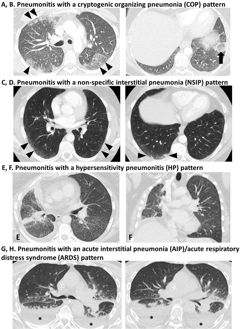
Spectrum of radiographic manifestations of PD-1 inhibitor-related pneumonitis.
A, B. Pneumonitis with a COP pattern in a 30-year-old female with Hodgkin lymphoma treated with nivolumab and ipilimumab combination therapy (Patient 15).
CT scan of the chest at 11.5 months of therapy demonstrated a development of ground-glass and reticular opacities and consolidations involving both lungs with a multifocal distribution (arrowheads, A), representing a COP pattern. One of the involved areas in the left lower lobe demonstrated a “reversed halo” sign (arrow, B) with central GGO surrounded by dense air-space consolidation of crescentic shape, which has been reported as a radiographic manifestation of COP.
C, D. Pneumonitis with a NSIP pattern in a 58-year-old male with melanoma treated with nivolumab monotherapy (Patient 1).
Chest CT scan at 1.7 months of therapy demonstrated new ground glass and reticular opacities and consolidations (arrowheads, C, D) indicative of a NSIP pattern.
E, F. Pneumonitis with a HP pattern in a 40-year-old female with lung adenocarcinoma treated with nivolumab monotherapy (Patient 12).
Chest CT scan at 1.2 months of therapy demonstrated new diffuse GGOs and centrilobular nodularity in both lungs, indicative of a HP pattern of pneumonitis. Note that the consolidations and interlobular septal thickening in the right lung demonstrate tumor involvement by lung cancer, which were present since the baseline scan.
G, H. Pneumonitis with an AIP/ARDS pattern in a 70-year-old man with melanoma treated with sequentially administered nivolumab and ipilimumab combination therapy (Patient 3).
Chest CT scan at 5.6 months of therapy demonstrated ground-glass and reticular opacities, consolidations and traction bronchiectasis as well as pleural effusions (asterisks), involving both lungs.