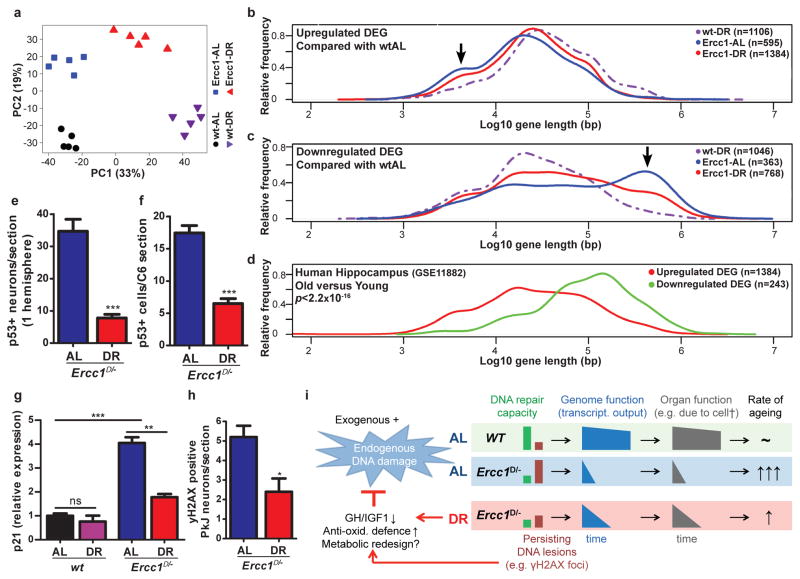
Figure 3. Dietary restriction in Ercc1Δ/− mice resembles wtDR and preserves genome function.
a, Principal component analysis (PCA) of full genome liver RNA expression profiles of 11-week old Ercc1Δ/− mice under AL (blue squares) and DR (red triangles) and wt controls (AL: black circles; DR: purple triangles). This analysis takes into account all the genes in the microarray platform. The two main principal components PC1 and PC2 explain 52% of the variability in the original dataset: PC1 (x-axis, 33%) differentiates on the basis of expression changes induced by DR, independent of genotype; PC2 (y-axis, 19%) reflects differences associated with genotype. b–c, Relative frequency plot of gene length (log scale) of DEGs in: wt-DR vs. wt-AL (purple interrupted lines), Ercc1Δ/− AL vs. wt-AL (blue lines) and Ercc1Δ/− DR vs. wt-AL (red lines). Plots are shown for only up-regulated genes (b), and only down-regulated genes (c). Black arrows indicate the extra peak of up-regulated short genes (b) and peak of down-regulated long genes (c) in Ercc1-AL. d, Relative frequency plot of gene length of DEGs in hippocampus of approximately 80 year old humans versus approximately 20 year young humans. Upregulated genes are depicted in red and downregulated genes in green. The DEGs from human hippocampus were selected using a log2-fold change cut-off of 0.5 and FDR <0.05. The dataset used corresponds to NCBI gene expression omnibus, number GSE11882. e–f, P53-positive cells were counted in neocortex of 3 consecutive transverse brain sections (e) at the level of the Bregma (Mouse brain atlas, Paxinos) and 3 consecutive C6 cervical spinal cord sections (f). Sections from 16-week old DR Ercc1Δ/− mice (n=4) show significantly reduced levels of P53-positive cells (p<0.0001 for neocortex and p=0.0002 for spinal cord) as compared to sections from AL mice (n=4). g, Relative expression changes in the P53 target gene p21 in 11-week old wt and Ercc1Δ/− mice by DR (n=5) h, yH2AX-positive Purkinje (PkJ) neurons were counted in cerebellum of 5 consecutive transverse brain sections in AL- and DR-treated Ercc1Δ/− mice (p=0.014). Error bars indicate mean ± SE. * p<0.05, ** p<0.01, ***p<0.001. i. Mechanistic model for the anti-aging effect of DR. With aging DNA damage from exogenous and endogenous (metabolic) sources accumulates, which is accelerated in repair-deficient Ercc1Δ/− mice. Stochastic DNA lesions reduce transcriptional output in a gene-size dependent manner leading to cell dysfunction and death, stem cell exhaustion, organ/tissue atrophy and functional decline, causing aging-related diseases. Accumulating DNA damage and DR trigger an anti-aging response, which involves suppression of growth, up-regulation of anti-oxidant defense and presumably metabolic redesign which reduces steady state levels of reactive metabolites, thereby preserving genome integrity and delaying aging-related functional decline.

