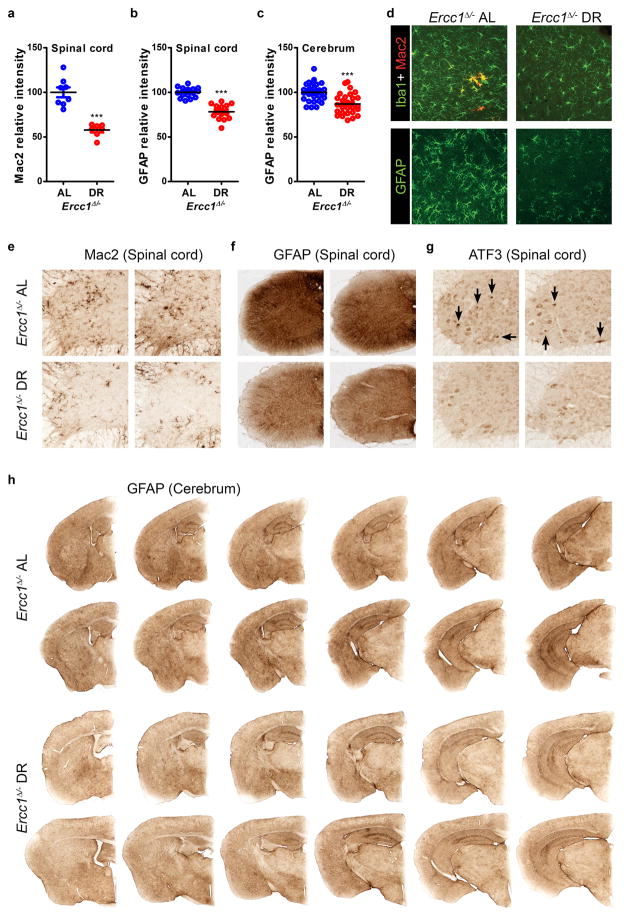
Extended Data Figure 5. Dietary restriction improves microgliosis and astrocytosis in brain and spinal cord of Ercc1Δ/− mice.
a–c, Quantification of the relative intensity of consecutive transverse brain and spinal cord sections immunoperoxidase-stained for Mac2 in spinal cord (a), and GFAP in spinal cord (b) and cerebrum (c). n>3 animals/group; bars indicate group medians. d, Iba1, Mac2, and GFAP immunofluorescent confocal images showing that reduced astrocytosis (GFAP) in cortex is paralleled by reduced staining for microglia (Iba1). Also Mac2-immunoreactivity, which outlines a subset of phagocytosing microglia cells, is reduced in 16 week old DR (n=4) as compared to AL (n=3) cortex in Ercc1Δ/− mice. e–g, Representative pictures of spinal cord sections of 16 weeks old AL and DR Ercc1Δ/− mice immunoperoxidase-stained for Mac2 (e) GFAP (f) reflecting reduced microgliosis and astrocytosis respectively, in the DR nervous system. Immunoperoxidase-stained spinal cord sections for ATF3 (g) showed that activation of the stress-inducible transcription factor ATF3 (which is induced following genotoxic stress via p53-dependent and -independent pathways) is less pronounced in DR nervous system. Per condition, sections from two different animals are presented next to each other. Black arrows indicate cells with high nuclear ATF3 staining. h, Representative pictures of consecutive transverse brain sections of 16 weeks old AL and DR Ercc1Δ/− mice immunoperoxidase-stained for GFAP, showing reduced GFAP staining, in the DR nervous system. Per animal, represented in one row, six 40μm slices are shown with 360μm cerebrum thickness in between each slice. Error bars denote mean ± SE. ***p<0.001.

