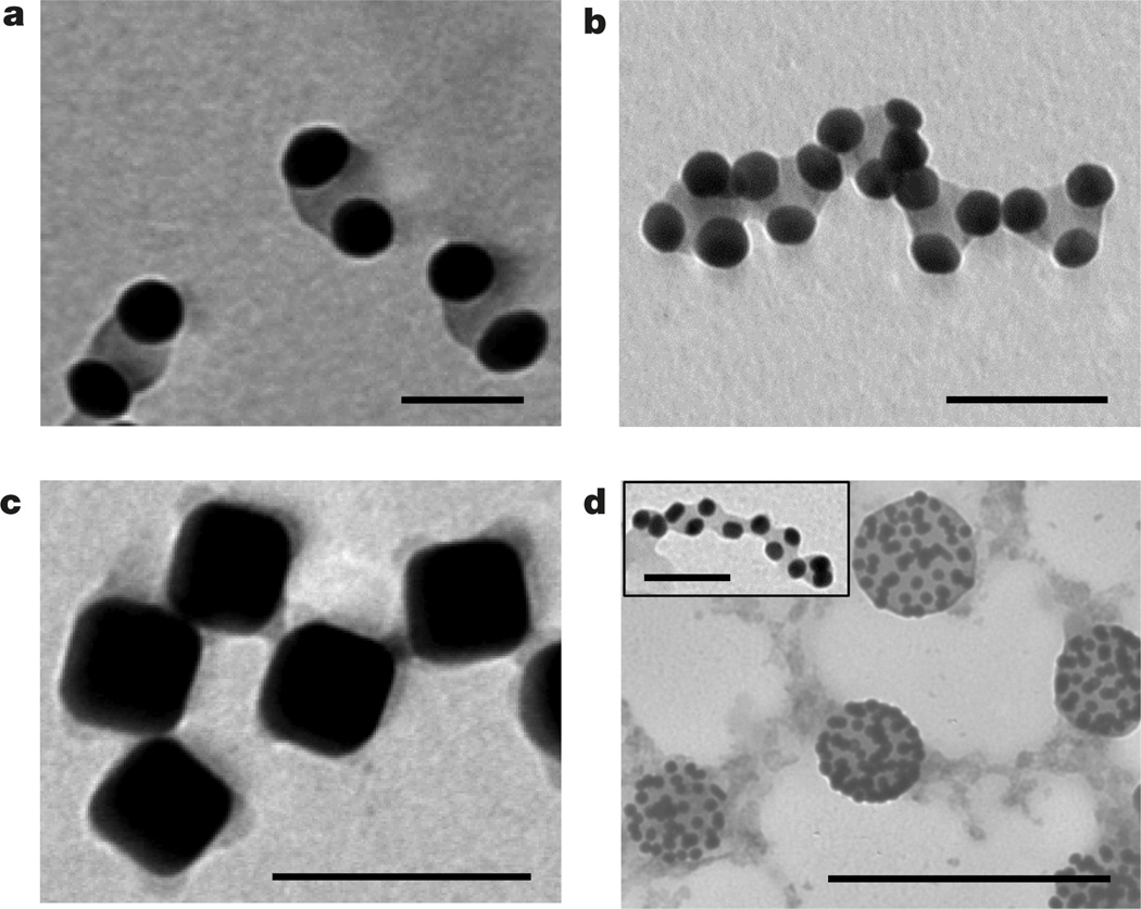
Figure 4. Self-assembly of patterned nanoparticles.
a, Dimers of single-patch gold nanospheres. b, Self-assembly of trimers of single-patch gold nanospheres in chains. In a and b the nanospheres were capped with polystyrene-50K and incubated for 15 days in the DMF/water solution at Cw = 4 vol% at 40 °C. Scale bars in a and b are 40 nm. c, Self-assembly of patchy silver nanocubes functionalized with polystyrene-50K in the DMF/water mixture at Cw = 20 vol%; scale bar is 100 nm. d, Self-assembly of gold nanospheres on the surface of droplets enriched with free nonthiolated polystyrene. The self-assembly was induced by adding water at Cw = 4 vol% to the mixed solution of free non-thiolated polystyrene (Mn = 50,000 g mol−1) and gold nanospheres tethered with polystyrene- 50K in DMF; scale bar is 250 nm. The inset to d shows self-assembly of patchy polystyrene-50K-capped gold nanospheres in the DMF/water mixture at Cw = 4 vol% in the presence of 0.625 nM of non-thiolated polystyrene, following 5 min sonication of the solution. Scale bar is 40 nm.

