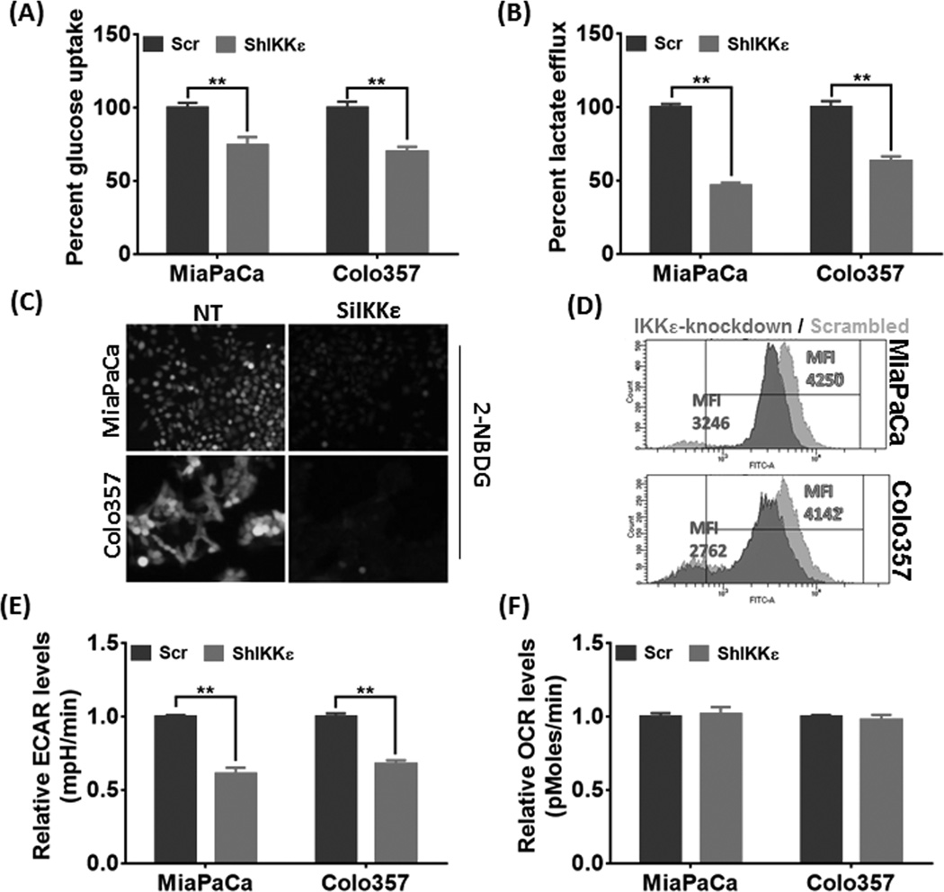
Figure 2. IKKε-silencing suppresses glucose-uptake and consumption in PDAC cells.
(A) Glucose-uptake and (B) lactate-efflux was measured in the used culture-media and normalized to cell counts. The data is depicted as percent change in IKKε-silenced cells relative to their respective controls. (C & D) PDAC cells were transfected with non-targeting control (NT) or IKBKE-targeting (SiIKKε) siRNAs. After 72 hours, cells were cultured in glucose-free FBS-free media for 6 hours and further incubated with glucose-free media supplemented with 100µM 2-NBDG for 3 hours. Thereafter, either the cells were visualized under fluorescent microscope and photographed (C), or subjected to flow cytometry analysis (D). Representative images are from independent experiments. (E & F) To examine basal ECAR and OCR, 4×10⁴ cells/well were seeded in XF24 cell-culture microplates and incubated at 37°C overnight. Next day, culture-media was replaced with XF Assay Medium (supplemented with 5mM Glucose) and plates loaded into the XF24 analyzer. Data is presented as mean ± S.D., n=3; **p≤0.05.

