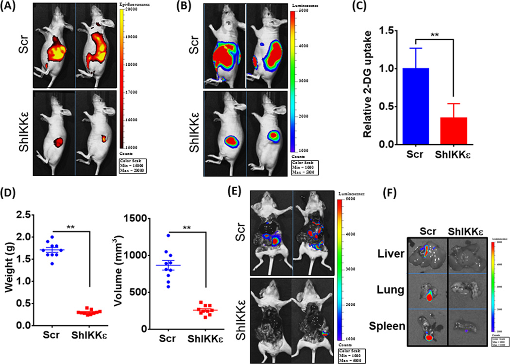
Figure 6. IKKε-downregulation suppresses glucose-uptake, tumor growth and metastasis in orthotopic PDAC xenografts.
Luciferase-tagged control or IKKε-silenced MiaPaCa cells were implanted into the pancreas of athymic nude mice (n=10 per group). (A) A day prior to end-point, mice were injected with fluorescent analog of glucose, 2-DG (100µL) intraperitoneally and epiflourescence measured after 24h using the IVIS imaging system. (B) Prior to sacrificing the mice, D-luciferin (150 mg kg−1 body weight) was injected intraperitoneally and bio-luminescence imaging data recorded using the IVIS imaging system. (C) In vivo glucose-uptake normalized with bioluminescent tumor measurements at the end time point and shown as relative 2-DG uptake. (D) After sacrifice, tumors were resected and measured for weight and volume, and (E) mice imaged to visualize metastases. (F) To further confirm metastases, livers, lungs and spleens were carefully removed from the mice and imaged separately. All data is representative. Bars represent the mean ± S.D. (n = 10 mice); **p ≤ 0.05.

