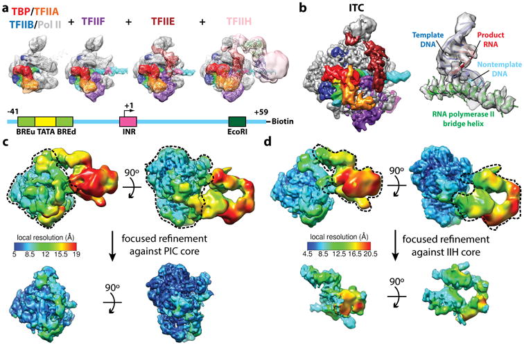
Figure 3. Structural determination of human TBP-based PIC.

(a) Reconstitution strategy for human PIC by sequential assembly. Color scheme for the components of the PIC is shown at the bottom. Cryo-EM reconstructions of PIC assembly intermediates for (from left to right): TBP–TFIIA–TFIIB–DNA–Pol II, plus TFIIF, plus TFIIE, and negative stain reconstruction of the holo PIC including TFIIH. Schematic of the DNA highlights the relative positions of the core promoter elements and restriction site (bottom). (b) Cryo-EM reconstruction of the core PIC in the ITC state (left) and near-atomic resolution details around the Pol II active site (right). (c,d) Refinement strategies for the “core” PIC in the CC (c) and the TFIIH core complex in the open promoter states (d). The local resolution estimation shows flexibility for TFIIH. Focused refinements (using the masks marked by dashed lines) improved alignment accuracy and thereby the resolution for targeted regions.
