Figure 2.
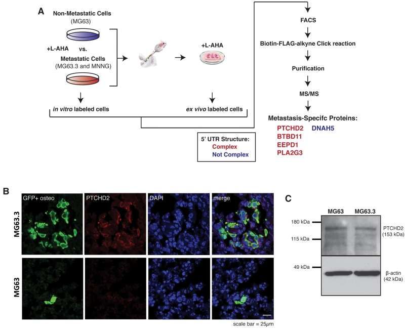
Proteins uniquely synthesized by highly metastatic osteosarcoma cells during colonization of the lung contain complex 5’ UTR structure.
A) Schematic of experimental design for proteomic identification of newly synthesized proteins in ex vivo lung culture by bioorthogonal non-canonical amino acid tagging (BONCAT). Highly metastatic (MG63.3 and MNNG) and non-metastatic (MG63) human osteosarcoma cells were grown in vitro and in ex vivo lung culture in methionine-deplete media supplemented with the methionine surrogate L-azidohomoalanine (L-AHA) to label newly synthesized proteins over 24 hour growth period. GFP+ cells were isolated by FACS, newly synthesized proteins were purified and detected by tandem mass spectrometry. Proteins uniquely identified in highly metastatic cells growing in ex vivo lung culture are listed. The structure and ΔG of the 5’ UTR of mRNA for these genes were predicted using the ViennaRNA Package 2.0 software(25). Genes with predicted 5’ UTR ΔG’s of −30 kcal/mol or less were considered complex/weakly translated.
B) In Vivo images of GFP+ non-metastatic (MG63) and highly metastatic (MG63.3) cells in the lung 7 days following tail-vein injection of 1×10⁶ cells. Sections stained for GFP, PTCHD2, and DAPI. Images were captured using a 63× lens on a Zeiss LSM-710 laser scanning confocal microscope.
C) Immunoblot analysis of PTCHD2 expression in non-metastatic (MG63) and highly metastatic (MG63.3) cells growing in in vitro culture. Images cropped to show relevant bands.

