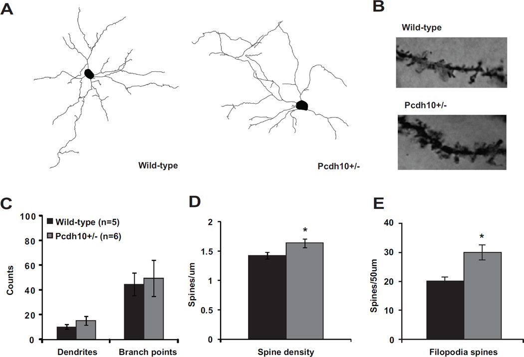
Figure 4. Increased filopodia-type spines on lateral/basolateral amygdala neurons of Pcdh10+/− males.
a) Representative dendritic reconstructions from lateral/basolateral (LA/BLA) amygdala neurons from wild-type and Pcdh10+/− males. b) Representative dendritic lengths from LA/BLA neurons from wild-type and Pcdh10+/− males. c) Counts of dendrites and branch points in LA/BLA of wild-type and Pcdh10+/− males. A mixed model was used to compare the mean dendrites and branch points between the two genotypes, while considering the non-independence of multiple measurements within some mice. There was no difference in dendrites (p=0.190) or branch points (p=0.771) between the genotypes. d) A mixed model was used to compare the mean spine density in the LA/BLA between the two genotypes, while considering the non-independence of multiple measurements within some mice. Based on the available literature on the role of Pcdh10 in spine elimination (17), a one-tailed test was applied to test our a priori directional hypothesis. Indeed, Pcdh10+/− mice had significantly higher spine density that WT littermates (p=0.048). e) A mixed model was fit separately for each spine type with a one-sided test, based on the a priori hypothesis of higher spine counts in Pcdh10+/− mice (17). Relative to WT, Pcdh10+/− amygdala neurons showed significantly higher levels of filopodia spines (p=0.030)

