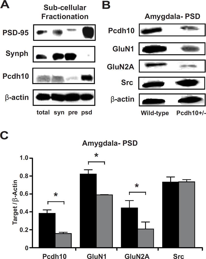
Figure 5. Decreased Pcdh10 in the PSD of Pcdh10+/− males was associated with decreased GLUN1 in the PSD.
a) Pcdh10 was found to be concentrated in the post-synaptic density fraction (psd) where PSD-95, the marker for PSD is most concentrated; whereas it was found to be poorly represented in presynaptic fraction (pre) where Synaptophysin (Synph), the presynaptic marker, is heavily concentrated. b) Representative blots of Pcdh10, GluN1, GluN2A and β-actin in the PSD fractions of wild-type and Pcdh10+/− mice showing decreased Pcdh10, GluN1, and GluN2A in the amygdala of Pcdh10+/− mice. c) Pcdh10+/− mice showed significantly reduced levels of Pcdh10 (p<0.001), GluN1 (p<0.001), and GluN2A (p=0.011), but not SRC (p=0.939).

