
Figure 2.
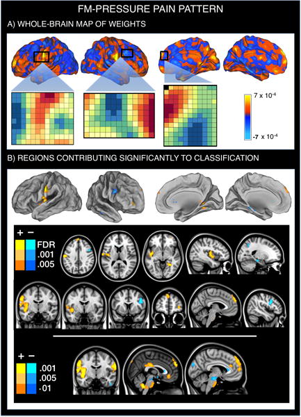
Multivariate brain pattern that predicts fibromyalgia status on the basis of brain activation during painful (pressure) stimulation. Positive weight values reflect higher pain-evoked activation in FM patients relative to healthy participants, whereas negative weight values reflect reduced pain-evoked activation in FM patients. A. SVM pattern of whole-brain voxel weights that optimizes classification of FM patients and healthy participants. We provide the voxel-by-voxel weights for three representative regions (anterior SII, right dorsolateral and dorsomedial PFC) to illustrate the concept of weighted pattern. B. Regions whose voxel weights contributed most reliably to the prediction of FM status (q<0.05 FDR-corrected for the first two rows; p-uncorrected<0.001 to further illustrate the findings).