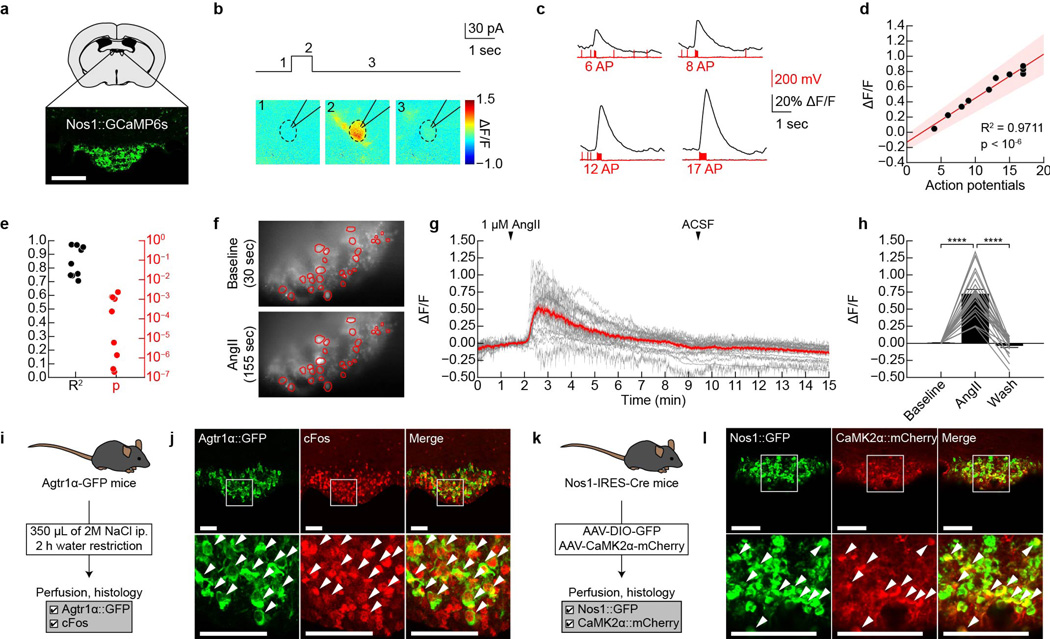
Extended Data Figure 2. GCaMP6s faithfully reports SFONos1 neuron activity in acute slices.
a, Expression of GCaMP6s in SFONos1 neurons from AAV5-EF1α-FLEX-GCaMP6s (scale bar, 100 µm). b, Representative fluorescence images of a neuron given a 30 pA current injection for 700 msec in acute slice (1 of 9 cells). c, Representative traces showing calcium responses in response to 30 pA current injections of increasing duration to produce increasing numbers of action potentials (1 of 9 cells). d, Relationship between number of action potentials and ΔF/F for the representative neuron in panel c (shaded area denotes 95% confidence interval). e, R2 and P-value for linear relationship between number of action potentials and ΔF/F (n = 9 cells). Panels f–j demonstrate that SFONos1 neurons are homogeneously responsive to both angiotensin and salt challenge. f, Representative fluorescence images showing SFONos1 neuron activity before and during bath application of angiotensin (1 of 3 experiments; red circles, identified neurons). g, 24/27 (~90%) identified SFONos1 neurons were activated by bath application of angiotensin (red line, mean; grey lines, individual activated neurons). h, Quantification (****P < 0.0001, two-way repeated-measures ANOVA, n = 24 activated neurons). i, Experimental design to test if a single population of SFO neurons is responsive to both angiotensin and salt challenge. j, Co-localization of Agtr1α::GFP and salt challenge-induced cFos indicates that SFONos1 neurons are homogeneously responsive to both angiotensin and salt challenge (scale bars, 100 µm). k, Experimental design to test if a SFO neurons express the excitatory neuron marker CaMK2α. l, Co-localization of CaMK2α::mCherry and Nos1::GFP indicates that SFONos1 neurons are excitatory (scale bar, 100 µm). Values are mean ± s.e.m. (error bars or shaded area).

