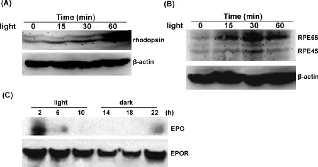
Fig. 1.
Light and circadian regulation of EPO/EPOR. Retinal cultures derived from rat or early passages of human RPE cells were exposed to light. Proteins were separated by SDS-PAGE and visualized by Western blot test. A: Rhodopsin was up-regulated in retinal cells after exposure to 5,000 lux light. B: RPE65 was increased after 15 min exposure to 5,000 lux light in the RPE. Truncation of RPE65 to 45 kDa was also seen at 30 min. C: Expression of EPO/EPOR in the 12 hr light/12 hr dark in vivo. Two hours’ exposure to room light (300 lux) rapidly increased the expression of EPO. Continuous exposure to light subsequently decreased EPO. Expression of EPO was recovered after prolonged incubation in the dark.

