Fig. 6.
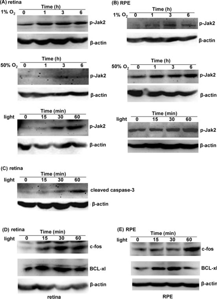
Downstream regulators of EPO in the retina and the RPE. Retinal cultures derived from rat or early passages of human RPE cells were exposed to 1% O2, 50% O2, or 5,000 lux light. Proteins were separated by SDS-PAGE and visualized by Western blot test. A: Expression of p-Jak2 increased in the retina exposed to 1% O2, 50% O2, or 5,000 lux light. B: Expression of p-Jak2 increased in the RPE exposed to 1% O2, 50% O2, or 5,000 lux light. C: Expression of activated caspase-3 after exposure to light in retinal cells. Until 30 min of light exposure, the level of cleaved caspase-3 in retinal cells was unchanged, indicating that endogenous neuroprotective or antiapoptotic proteins including EPO/EPOR against light stress were up-regulated. At 60 min, increased level of cleaved caspase-3 was compatible with significant cell death. Expression of c-fos and BCL-xl in the retina (D) and RPE cells (E). These antiapoptotic proteins were up-regulated in the light.

