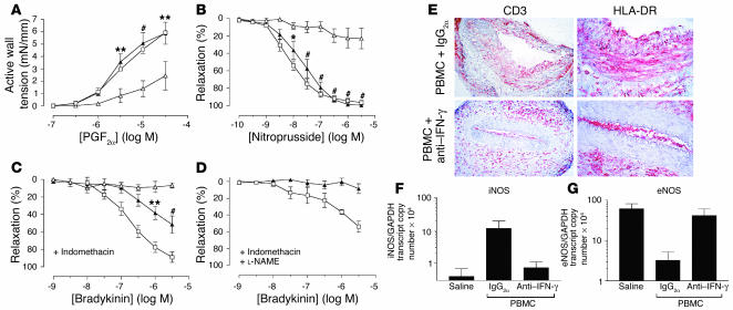
Figure 3.
Effects of IFN-γ neutralization on T cell–mediated graft dysfunction in vivo. Transplanted human arterial segments were recovered from mice injected with saline alone (open squares) or PBMCs together with anti–IFN-γ mAb (filled triangles) or control mAb (IgG2a) (open triangles) for 2 weeks (n = 5 matched triplicates from three experiments). (A and B) Endothelium-independent responses to various concentrations of PGF2α (A) and nitroprusside (B). (C and D) Endothelium-dependent response to bradykinin before (C) and after (D) treatment with L-NAME. *P < 0.05; **P < 0.01; #P < 0.001 vs. PBMC + IgG2a group, n = 3–5. (E) Immunohistochemistry of graft sections stained for human CD3 and HLA-DR. The luminal HLA-DR–positive cells are also human CD31–positive (data not shown). Goat anti-mouse secondary Ab alone did not stain the grafts. Original magnification: CD3 panels, ×200; HLA-DR panels, ×400. (F and G) RNA transcript levels of iNOS (F) and eNOS (G) in whole tissue sections analyzed by quantitative RT-PCR.

