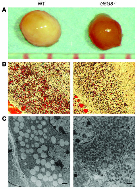
Figure 1.
Comparison of the adrenal glands from female G5G8–/– mice with those from female wild-type mice. (A) Adrenal glands from 6-month-old female wild-type and G5G8–/– mice. (B) Sections from the adrenal cortex of a female wild-type mouse and a female G5G8–/– mouse after staining with Oil-Red O (×20 magnification). (C) Electron micrographs of the adrenal zona fasciculata of wild-type and G5G8–/– mice. Adrenal glands were removed from 6-month-old female wild-type and G5G8–/– mice, and electron microscopic sections were prepared and examined as described in Methods. Electron micrographs were taken at a magnification of ×2,500. Scale bars: 1 μm.

