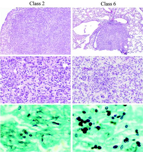
FIG. 2.
Hematoxylin and eosin staining of lung tissue. Chronic inflammation was characteristic of class 2 infection (top panel), consisting of macrophages, lymphocytes, fibroblasts, and rare granulocytes but no necrosis (middle panel). H. capsulatum yeast forms were scattered diffusely in the Gomori methenamine silver stain (bottom panel). Lungs of mice infected with class 6 showed immature granuloma (top panel) with prominent early caseation necrosis and numerous macrophages and neutrophils (middle panel), and GMS stain shows yeast forms confined focally within granuloma (bottom panel). Magnifications of top, middle, and bottom panels, ×21.75, × 108.75, and ×435, respectively (original magnifications, ×25, ×125, and ×500, respectively).

