FIG.1.
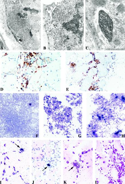
Histological and virological features of muscle tissues and cultures. Electron micrographs of deltoid muscle sections from patient CB showing intracytoplasmic accumulation of abnormal filaments (magnification, ×9,800) (A) and clusters of straight (B) and paired helical (C) filaments seen at higher magnification (×14,000 and ×18,000). Immunochemical detection of CD4+ (D) and CD8+ (E) T cells on muscle sections counterstained with Harris hematoxylin. Detection of tax mRNA-positive cells among fresh PBMCs (F) and CD4+ T lymphocytes sorted from muscle culture (G) compared to PHA-stimulated CD4+ T cells from blood (H) (counterstained with Giemsa; exposure time, 8 days). In situ detection of tax mRNA with an antisense probe on muscle sections from the first (I and J) and the second (K) biopsies; arrows show focal positivity for HTLV-1 in mononuclear infiltrated cells and not in muscle fibers. Negative control analyses were performed with a tissue section from the second biopsy with a sense probe (L). Sections were counterstained with hematoxylin-eosin, and the exposure time was 21 days.

