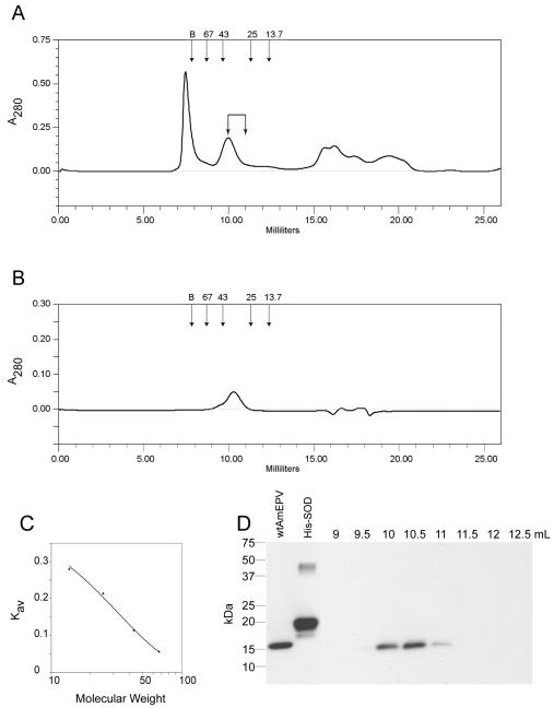
FIG. 6.
Size determination of AMVSOD from infected cells. (A) Chromatogram from a Superdex 75 column of a mixture of proteins from wild-type AmEPV-infected cells. Arrows at the top indicate the locations of peaks for each size standard. Elution volume is indicated on the x axis. (B) Chromatogram of bovine SOD for comparison. The calculated size is 34.6 kDa. (C) The standard curve for the column is derived from proteins of known size: albumin, 67 kDa; ovalbumin, 43 kDa; chymotrypsinogen A, 25 kDa; and RNase A, 13.7 kDa. B indicates the position of the blue dextran peak used to determine the void volume of the column. (D) Western blot of 0.5-ml fractions from the column with monoclonal antibody 2B8-1C9 to detect AMVSOD. Lane 1, total protein; lane 2, 0.25 μg of purified His-SOD, lanes 3 to 10 are labeled according to the starting elution volume of the fraction and correspond to fractions 19 to 26, respectively. AMVSOD elutes predominantly at 10 to 11 ml.

