FIG. 1.
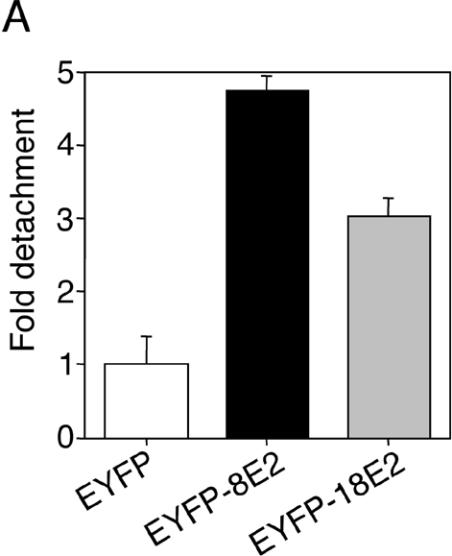
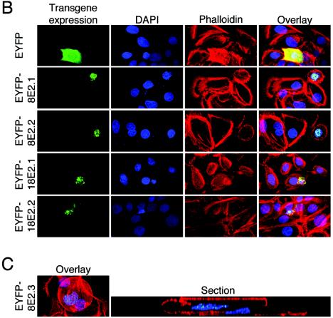
(A) Detachment of HPV E2-expressing keratinocytes from culture dish. Primary human keratinocytes were seeded in 12-well plates and transfected with 0.77 μg of pEYFP-C1, pEYFP-HPV8-E2, or pEYFP-HPV18-E2 expression construct. After 48 h, the number of fluorescent cells floating in the supernatants was counted. The data were normalized to the transfection efficiencies in each group. Shown is the relative detachment compared with the EYFP control in two independent experiments. Error bars indicate standard deviations. (B) HPV E2-expressing keratinocytes display distinct morphological features. Keratinocytes seeded on glass coverslips were transfected as described above. After 48 h, cells were fixed, permeabilized, and stained with AlexaFluor-633-phalloidin (red fluorescence) and DAPI (blue fluorescence). EYFP- or EYFP-E2-expressing cells (two independent stainings) (green fluorescence) were analyzed by confocal microscopy. Overlays are shown in the right panels. (C) HPV E2-expressing keratinocytes can be found on top of nontransfected cells. Keratinocytes transfected with pEYFP-HPV8-E2 and stained as described above were analyzed by confocal microscopy (left panel). A section (gray line) through a nontransfected cell (right cell, nucleus in blue) and an EYFP-HPV8 E2-expressing cell lying above (left cell, nucleus in blue with EYFP-fluorescence in green) is shown in the right panel.


