FIG. 3.
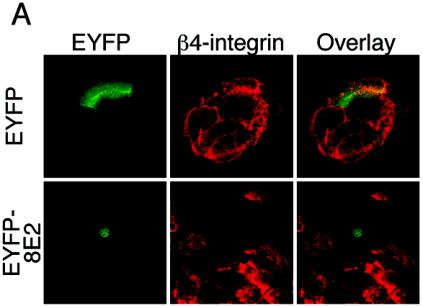
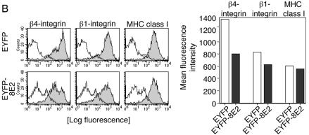
(A) Analysis of the β4-integrin expression pattern by confocal laser scanning microscopy. Keratinocytes seeded on glass coverslips were transfected with pEYFP-C1 vector or the pEYFP-HPV8-E2 or pEYFP-HPV18-E2 expression construct. After 48 h, cells were fixed, permeabilized, and stained with anti-β4-integrin (red fluorescence) primary and Cy3-conjugated secondary antibodies. EYFP-expressing cells (green fluorescence) were analyzed by confocal microscopy. Overlays are shown in the right panel. All pictures of β4-integrin stainings were recorded at the focal plane corresponding to the cell-substrate interface. (B) HPV8 E2 strongly down-regulates β4-integrin compared with β1-integrin and MHC class I expression levels. Keratinocytes were seeded onto plastic dishes. After 24 h, they were transfected with expression constructs encoding EYFP or EYFP-tagged HPV8 E2. Forty-eight hours later, cells were harvested, stained, and gated for EYFP fluorescence. Expression levels of β4- and β1-integrins as well as MHC class I were determined by flow cytometry and are represented by solid gray histograms (left panels). The white histograms represent isotype-matched controls (left panels). Mean fluorescence intensities of the respective stainings are shown in the right panel.


