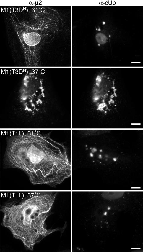
FIG. 7.
Distribution of cUb and T3DN or T1L μ2 in transfected cells at 31 or 37°C. CV-1 cells were transfected with 2 μg of pCI-M1(T3DN) (top two rows) or 2 μg of pCI-M1(T1L) (bottom two rows) per well. Transfected cells were incubated at 31 or 37°C as indicated. At 18 h p.t., cells were fixed and immunostained with rabbit anti-μ2 polyclonal serum followed by anti-rabbit IgG conjugated to Alexa 594 (α-μ2) (left column) and with mouse MAb FK2 against cUb followed by anti-mouse IgG conjugated to Alexa 488 (α-cUb) (right column). Bars, 10 μm.

