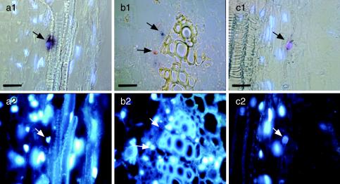
FIG. 5.
In situ detection discriminating between TYLCSV and TYLCV in N. benthamiana tissues from stems of doubly infected N. benthamiana, hybridized with a biotin-labeled TYLCSV probe staining red (b) or a DIG-labeled TYLCV probe staining blue (a and b). Violet color (c) indicates the presence of both viruses. Samples were examined by either combined DIC and epifluorescence microscopy (a1 and c1), DIC microscopy (b1), or epifluorescence microscopy alone to visualize DAPI-stained nuclei (a2, b2, and c2). Bar, 25 μm. Arrows point at the same sites in the corresponding pictures. Note the predominantly TYLCV- or TYLCSV-infected nuclei in neighboring cells (b1) and the presence of both viruses within one nucleus (c1).

