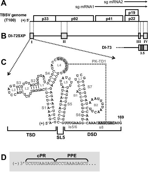
FIG. 1.
TBSV genome and relevant viral RNAs. (A) TBSV RNA genome. The genome is represented by a thick black line with coding regions depicted as boxes, and approximate molecular masses (in thousands) of the encoded proteins are shown, prefixed with “p.” (10). Two subgenomic mRNAs (sg mRNA1 and sg mRNA2) produced during infections are shown as arrows above the genome. (B) Prototypical TBSV DI RNAs DI-72SXP and DI-73. Shaded boxes represent TBSV genomic segments present in DI RNA, whereas black lines represent genomic segments that are absent. DI-73 is similar to DI-72SXP in that it contains RI through RIV (RI and RII are not shown). However, the former has an extra 3′-proximal sequence, region 3.5 (dark grey box), that is absent in the latter. (C) RNA secondary structure model for the TBSV 5′ UTR. The TBSV TSD, SL5, and the DSD are indicated by brackets below the structure (23, 32). Sequences forming a TSD-DSD pseudoknot (PK-TD1) are shaded in grey. RNA stem and loop structures are labeled, and TBSV genome coordinates are provided. is5/6, intervening sequence 5/6. (D) Sequences of the TBSV promoter (cPR) and enhancer (PPE) for positive-strand RNA synthesis located at the 3′ terminus of the DI RNA negative strand.

