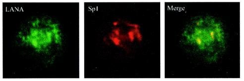
FIG. 3.
Immunocolocalization of LANA and Sp1 from BC-3 cells. BC-3 cells were fixed in methanol-acetone (1:1) and incubated with an anti-LANA serum. LANA was localized by use of an Alexa fluor-conjugated secondary antibody. Sp1 was localized in the same cells by use of a mouse monoclonal anti-Sp1 antibody and a Texas red-conjugated secondary antibody. The merged image shows the colocalization of these two proteins in the nuclear clusters, where the predominant amount of the Sp1 signal was observed. LANA staining was observed in a somewhat speckled, fibrous pattern with additional signals in the nuclear clusters dominated by Sp1 staining.

