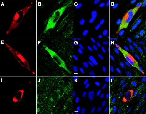
FIG. 4.
Localization of several packaged RNAs in infected fibroblasts during HCMV assembly. Fibroblasts were infected at a multiplicity of infection of 0.01 PFU/ml and processed for combined immunofluorescence and in situ hybridization at 72 h postinfection. In situ hybridization was completed with probes to UL21.5 (A to D), UL83 (E to H), and β-actin (I to L), shown in green. Immunofluorescence was completed with an antibody to the tegument protein pp28 (A, E, and I), shown in red. DNA was counterstained and appears blue (C, G, and K). Merged multicolor images (D, H, and L) and bars representing 10 μm (C, G, and K) are also shown.

