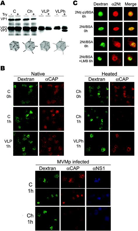
FIG. 3.
Nuclear export activity of 2Nt in transformed cells. (A) Heat-triggered exposure of 2Nt in MVM particles. Purified native empty capsids (C) and VLPs or the respective particles heated at 50°C for 10 min (Ch and VLPh) were tested for 2Nt exposure by trypsin (Try) digestion (23), and the cleavage of VP2 to form VP3 was visualized by immunoblotting with an α-VP antiserum. The four types of microinjected particles, with phosphorylated serine residues of 2Nt in natural capsids (black balls) or no phosphorylation in VLPs, are illustrated at the bottom. (B) Subcellular localization of MVM particles injected inside the 324K nucleus. Viral particles were injected with dextran-FITC in noninfected (upper panels) or infected (lower panels) cells and analyzed either immediately (0 h) or at 1 h postinjection by confocal laser microscopy after staining with the α-CAP serum and the α-NS1 MAb antibody where indicated. Shown are representative fields at different magnifications with clusters of injected cells and FITC-dextran marking intact nuclear membranes. (C) Confocal analysis of 2Nt(−p)-BSA and 2Nt-BSA protein conjugates microinjected into the 324K nucleus. Cells were fixed at the indicated times postinjection and stained with the α-2Nt antibody. Where indicated (+LMB), LMB (100 ng/ml) was added to the cultures after injection.

