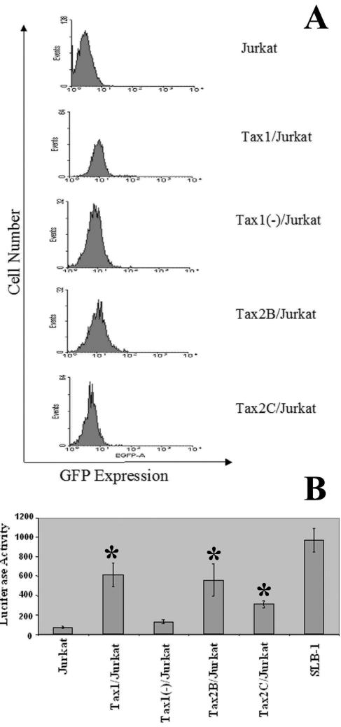
FIG. 1.
Characterization of Jurkat cell lines constitutively expressing Tax1 and Tax2. (A) Flow cytometric analysis of GFP expression. Jurkat cells were infected with LVs (MOI = 3), as previously described (62). Cells were serially diluted and clonally plated in 96-well plates at 48 h postinfection. After expansion, clones were randomly selected and analyzed for GFP expression by flow cytometry. GFP expression was assayed at 1, 6, and 12 weeks after clonal expansion, and GFP expression measured at 12 weeks is shown. The data were analyzed by using WinMDI 2.8 software. The clonal cell lines and LVs used for infection are as follows: Tax1/Jurkat, HR′CMV-Tax1/GFP; Tax1(−)/Jurkat, HR′CMV-Tax1(−)/GFP; and Tax2B/Jurkat and Tax2C/Jurkat, HR′CMV-Tax2/GFP. (B) Luciferase activity resulting from transfection of Jurkat clones with HTLV-1-LTR-Luc. Jurkat cell lines (107) were transfected with HTLV-1-LTR-Luc (24 μg; a gift from Kuan-Teh Jeang, National Institutes of Health, Bethesda Md.) by using Lipofectamine 2000 (Invitrogen). Cells were lysed in cell culture lysis reagent (Promega), and the luciferase activity was normalized for the amount of protein in each extract, as determined by a Bradford assay. SLB-1 is an HTLV-1-transformed T-cell line and contains multiple proviral integrations of HTLV-1. The experiment was performed three times, and the luciferase assays were carried out in triplicate. Error bars represent one standard deviation, and statistical analysis was performed by using the Student t test (P < 0.05), with Tax1(−)/Jurkat cells as a control. The transfection efficiencies ranged from 80 to 95% in SLB-1 cells as determined by GFP expression after cotransfection of a GFP reporter gene construct and HTLV-1-LTR-Luc.

