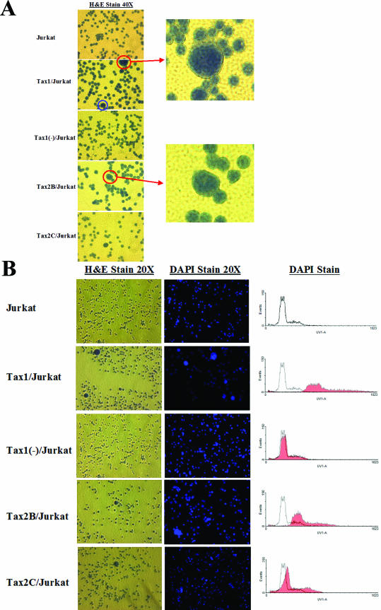
FIG.3.
Multinucleation in Tax1/Jurkat and Tax2/Jurkat cell cultures. LV transduced Jurkat cells (105) were plated onto poly-l-lysine-coated chamber slides, incubated in 1:1 methanol-acetone, and stained with hematoxylin for 2 min and eosin for 1 min (H&E). Cell lines were also stained separately with DAPI, a DNA-specific stain, for 10 min at room temperature. Photographs were taken with a SPOT digital microscope camera (Diagnostic Instruments, Inc., Sterling Heights, Mich.) on a Nikon Eclipse microscope. (A) The presence of multinucleated cells was detected by H&E staining (magnification, ×40). The blue circle represents a cell undergoing mitosis for reference. The red circles represent large, multilobulated cells representing ∼1% of total cells in the Tax1/Jurkat, Tax2B/Jurkat, and Tax2C/Jurkat cell cultures. Each enlarged field is photographed at ×80. (B) Hematoxylin-and-eosin (H&E)- and DAPI-stained Jurkat cell lines. Aliquots of cells (106) were stained with H&E (left panels) or DAPI (middle panels) and observed by microscopy (magnification, ×20). DAPI-stained cells were also analyzed by flow cytometry (Becton Dickinson). The shaded overlay represents LV-transduced Jurkat cells stained with DAPI compared to the parental Jurkat cell line stained with DAPI (right panels).

