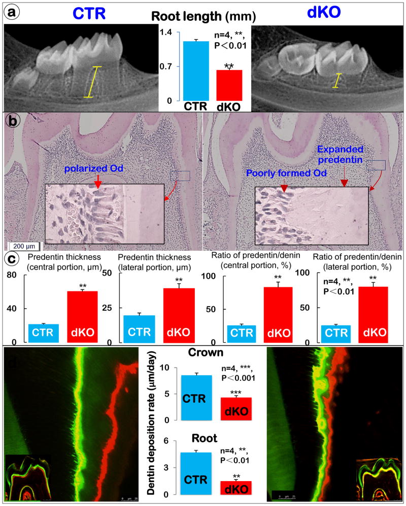
Figure 1. BMP1/TLL1 dKO mice display short roots and a dentinogenesis imperfecta-like phenotype (right panels).
a) X-ray examination of dKO mice revealed short molar root length. (n=4, **, P<0.01) b) H&E staining displayed some typical dentinogenesis imperfecta-like phenotype, including enlarged pulp with less pulp cells, expanded predentin, and a lack of odontoblast polarization. c) Quantitative analysis of the predentin thickness and predentin/dentin ratio in different root areas. d) Double fluorescence labeling of mandible from 3-week-old control (CTR) and dKO mice. The first injection (calcein) gave rise to a green label, whereas the second injection (Alizarin Red) produced a red line. The distance between the green and red labeling indicated the mineral deposition rate in the period between the two injections (5 days). The quantitative measurement of the distance between the two injections revealed a significantly lower mineral deposition rate both in the crown (moderate) and root dentin (severe) of dKO mice compared with the CTR group. (n=4, ***, P< 0.001, **, P< 0.01,).

