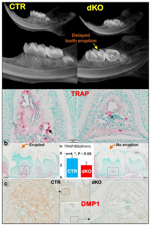
Figure 3. The dKO mice developed delayed molar eruption (right panels).
a) X-ray images showed a lack of molar eruption in the dKO mice compared to the age-matched controls, in which both 1st and 2nd molars are fully erupted; b) TRAP stain images showed more positive osteoclasts (black arrow) in CTR mice than in dKO mice, with a statistical difference between these two groups (n=4, *; P<0.05); and c) DMP1 immunostain images displayed a major defect in alveolar bone formation in the dKO mice.

