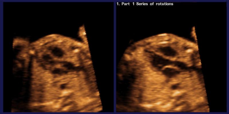
Figure 2.
Spatiotemporal image correlation (STIC) volume dataset of the fetal heart showing the left ventricular outflow tract view through the Fetal Intelligent Navigation Echocardiography (FINE) method. The left ventricular outflow tract was not successfully obtained using the diagnostic plane (left image). However, after Virtual Intelligent Sonographer Assistance (VIS-Assistance®) was activated, the automatic navigational movements allowed the left ventricular outflow tract to be successfully obtained (right image). Also see videoclip S3.

